
Most Popular


![[KH Explains] How should Korea adjust its trade defenses against Chinese EVs?](http://res.heraldm.com/phpwas/restmb_idxmake.php?idx=644&simg=/content/image/2024/04/15/20240415050562_0.jpg&u=20240415144419)


[Health-tech Korea] Tomocube’s light microscope enables realtime, 3-D observation of live cells
By Sohn Ji-youngPublished : March 5, 2017 - 16:46
The Korea Herald is publishing a series of articles highlighting South Korea’s promising start-ups in the emerging sectors of digital health care and next-generation medical devices. This is the third installment. – Ed.
DAEJEON — A medical device startup born from an applied physics lab at the Korea Advanced Institute of Science and Technology is out to introduce a next-generation 3-D microscope that uses light to observe living cells in real time.
Founded in 2015, Tomocube has developed a new holographic microscope that shoots weak laser light through samples of live cells and tissues. It uses their refractive index — how much the light bends, or refracts, when passing through a medium — to construct a three-dimensional image of their structure and internal properties.
The technique does not require any physical “labeling” or “staining” of the cells. Existing devices require injecting a fluorescent material into the cells and shining a strong light through them -- a process that often kills the cells or alters their original state.
“We expect our device to become highly useful in fields like cell biology, immunology and hematology as well as new drug development and medical diagnoses,” Tomocube’s co-founder and Chief Technology Officer Park Yong-keun told The Korea Herald.
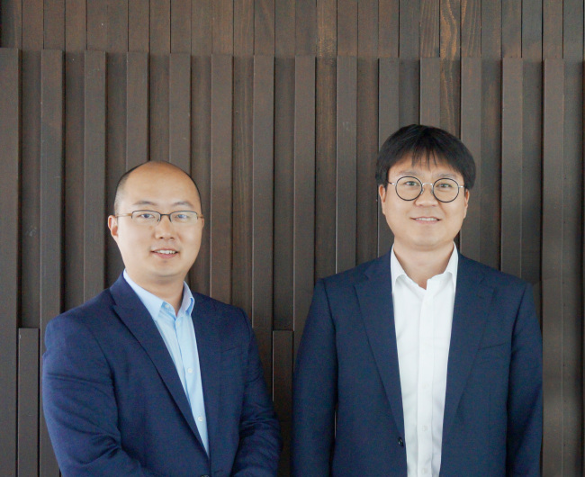
Park, who holds a Ph.D. in medical engineering from Harvard University, has dedicated much of his academic career researching photoptics. After joining KAIST’s physics department, Park built the initial prototype of its light microscope. He decided to later commercialize the device, establishing Tomocube.
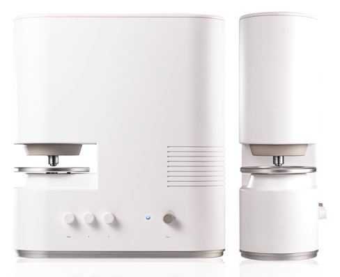
Tomocube’s “label-free” microscope, the HT-1, uses a self-coined technology called “holotomography,” which merges holography -- the photographic recording of a light field -- with computed tomography, or CT technology.
Just as a CT machine emits beams of X-rays through a patient’s body by revolving at a 360 degree angle, Tomocube’s device shines laser beams through a live cell or tissue at a 360 angle.
The HT-1 calculates the refractive values of these light beams to construct a 3-D image of these cells. It can also determine structural and chemical information, including their mass, morphology, protein concentrations and changes within the cellular membrane.
Most importantly, Tomocube’s light microscope lets scientists monitor how a cell changes over time -- whether on its own or in reaction to a foreign substance -- by conducting repeated light scans at fixed intervals.
“By using light, we can calculate a cell’s mass and track its internal dynamics by monitoring how different substances within a cell change over time,” Tomocube co-founder and CEO Hong Ki-hyun explained.
“This type of data is particularly important and useful when observing interactions between two cells, or between a cell and a substance (such as a drug),” Hong said.
For instance, Tomocube’s device could be used to discern the effects of a new drug on a patient’s liver. It could be used to analyze how fat within the cell decreases in reaction to the drug over time, he explained.
Because oil and water have differing masses, their refractive indexes also differ. By tracking the changes in the masses of the fat molecules inside a cell, one can discern how a drug is affecting its target.
A large pool of data on how a particular cell changes over the course of time -- when combined with image recognition capabilities powered by deep learning -- is also expected to help researchers detect early signs of diseases such as cancer, Hong said.
The HT-1’s ability to scan live cells with the same amount of light at constant intervals could help automate the cell observation process as well, he added.
Tomocube’s technology has already caught the attention of investors. The startup has attracted more than 4 billion won ($3.46 million) from Softbank Ventures Korea, Hanmi Science, the parent company of Korean drugmaker Hanmi Pharmaceutical and InterVest.
As of now, Tomocube has sold or freely provided for research purposes some dozen HT-1 units to universities and hospitals in Korea and abroad. It is now working to obtain the needed certification from multiple regulatory bodies to commercialize its light microscope in the US and Europe.
In doing so, CTO Park has already published more than 60 research papers detailing research studies conducted using the HT-1 to build up a pool of use cases for its new device.
As it prepares to expand sales, Tomocube expressed confidence in the competitive pricing of the HT-1, which has adopted a significantly cheaper price than that of existing devices in the global market.
Conventional light microscopes that use “staining,” sold by international medical device giants like Olympus, Nikon and Carl Zeiss are currently priced at between 200 million won to 500 million won, the startup said.
“Until now, these high-priced devices were primarily used at research labs as they were the best option available in the market,” said Hong. “We’re hoping to break this with an alternative, cost-efficient device.”
The company’s proposed target for this year is to sell at least 100 devices and record around 3 billion won in revenue, according to Hong.
“Our goal is to rise up as the world’s No. 1 holotomography company. We hope to become a notable example of a high-tech medical device company hailing from Korea,” the two co-founders said.
By Sohn Ji-young (jys@heraldcorp.com)


![[Today’s K-pop] Stray Kids to return soon: report](http://res.heraldm.com/phpwas/restmb_idxmake.php?idx=642&simg=/content/image/2024/04/16/20240416050713_0.jpg&u=)