

Most Popular


-
6
[Graphic News] More Koreans say they plan long-distance trips this year
![[Graphic News] More Koreans say they plan long-distance trips this year](//res.heraldm.com/phpwas/restmb_idxmake.php?idx=644&simg=/content/image/2024/04/17/20240417050828_0.gif&u=)
-
7
[KH Explains] Hyundai's full hybrid edge to pay off amid slow transition to pure EVs
![[KH Explains] Hyundai's full hybrid edge to pay off amid slow transition to pure EVs](//res.heraldm.com/phpwas/restmb_idxmake.php?idx=644&simg=/content/image/2024/04/18/20240418050645_0.jpg&u=20240419100350)
-
8
North Korea removes streetlights along cross-border roads with South

-
9
Russia's denial of entry of S. Korean national unrelated to bilateral ties: Seoul official

-
10
Farming households dip below 1m for first time in 2023

![[Graphic News] More Koreans say they plan long-distance trips this year](http://res.heraldm.com/phpwas/restmb_idxmake.php?idx=644&simg=/content/image/2024/04/17/20240417050828_0.gif&u=)
![[KH Explains] Hyundai's full hybrid edge to pay off amid slow transition to pure EVs](http://res.heraldm.com/phpwas/restmb_idxmake.php?idx=644&simg=/content/image/2024/04/18/20240418050645_0.jpg&u=20240419100350)


Scientists produce retina structure from blood-derived cells
By Yoon Min-sikPublished : March 14, 2012 - 18:08
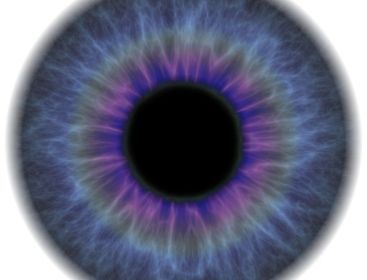
U.S. scientists have succeeded in making early retina structures by using stem cells from blood, marking a breakthrough toward treating eye diseases, Science Daily reported Tuesday.
The new findings can help study degenerative retinal disorder such as retinitis pigmentosa, a prominent cause of blindness in children and young adults, according to a statement by the University of Wisconsin- Madison research team.
Last year, the group led by Doctor David Gamm was able to create the most primitive structure of a retina with photoreceptors by using embryonic stem cells and stem cells from human skin. But the structures lacked the organization of a more mature retina.
This time, Gamm’s team used induced pluripotent stem cells (iPS) derived from blood gathered from donors. Induced pluripotent stem cell refer to a cell which can develop into any fetal or adult cell type but is free from ethnic debate because it does not require human ovum to produce.
Scientists extracted a type of blood cell called a T-lymphocyte, which is related to immunity, and reprogrammed the cells into iPS cells. Then they grew retina-like tissues from the iPS cells.
About 16 percent of the initial retinal structures developed distinct layers, which is a significant advance, as retina forms layer in normal human development.
The arrangement of layers was similar to what is found in the back of the eye.
Furthermore, the newly created cells possessed the ability to communicate information.
These facts suggest the potential to grow more complex retinal issues in a lab, by merely taking a blood sample from a patient.
“We don’t know how far this technology will take us, but the fact that we are able to grow a rudimentary retina structure from a patient’s blood cells is encouraging, not only because it confirms our earlier work using human skin cells, but also because blood as a starting source is convenient to obtain,” says Dr. David Gamm, the senior author of the study. “This is a solid step forward.”
The laboratory-built human retinal tissues can be used in various ways, including in drug-testing to ultimately replacing multiple layers of damaged retinal tissues.
Gamm said he hopes that the results from his team’s research can help people suffering from eye diseases in the future.
By Yoon Min-sik
<관련 한글 기사>
혈액에서 나온 세포로 망막 형성 성공!
미국 연구진이 혈액에서 추출한 세포를 이용해 망막의 초기 조직을 형성하는데 성공했다고 사이언스 데일리가 13일(현지시간) 보도했다.
이 발견은 어린이와 젊은 성인들 사이에서 나타나는 실명의 주된 원인인 망막색소변성증 (retinitis pigmentosa) 등 망막관련 질병 연구에 도움이 될 수 있다고 위스콘신-매디슨 대학 연구진은 밝혔다.
작년에, 데이비드 감 박사가 이끄는 연구진은 배아줄기세포(mbryotic stem cell)와 인간의 피부에서 추출한 줄기세포를 이용해 광수용기 (photoreceptor)을 가진 원시적인 형태의 망막조직을 만드는데 성공했다.
그러나 이 조직에서는 더욱 성숙한 형태의 망막에서 나타나는 구조가 발견되지 않았다.
이번 연구에서 감의 팀은 유도만능줄기세포 (induced pluripotent stem cells: iPS)를 혈액에서 추출해 이용했다. iPS 어떠한 세포로도 분화가 가능하지만 난자나 수정란을 이용하지 않아 윤리문제에서 자유로운 세포를 일컫는다.
과학자들은 혈액에서 면역력과 관련된 T림프구라고 불리는 세포를 꺼내 iPS로 변형시켰다. 그런 다음 iPS가 망막세포로 자라나도록 유도했다.
최초의 망막조직 중 16 퍼센트 정도에 뚜렷한 층이 생겨났는데, 인간의 안구에서도 층이 나타나기 때문에 이는 의미가 깊다고 할 수 있다.
또한 층의 구조 역시 안구의 뒷부분의 형태와 같았다.
그리고 새롭게 형성된 세포는 상호간에 정보를 전달할 수 있는 능력까지 갖췄다.
이러한 결과들은 차후에 환자의 혈액을 뽑는 것만으로 더욱 복잡한 구조의 망막조직을 배양할 수 있다는 가능성을 보여준다.
“이 기술이 어디까지 우리를 멀리 데려가 줄지 모릅니다만, 우리가 환자의 혈액을 이용해 기본적인 망막 조직을 배양했다는 사실은 고무적입니다. 우리가 기존에 인간의 피부세포를 이용하여 한 연구를 확고히 했을 뿐 아니라, 혈액은 쉽게 얻을 수 있기 때문에 의미가 깊습니다,”라고 감 박사는 밝히며 이는 의미 있는 발걸음이라고 말했다.
연구실에서 배양한 망막조직은 신약 시험 등 여러 가지로 활용될 수 있으며 최종적으로는 크게 손상된 망막을 대체하는데 쓰일 가능성도 있다.
감은 그의 연구가 안구관련 질병을 앓고 있는 사람들을 돕기를 희망한다고 말했다.


![[KH Explains] Hyundai's full hybrid edge to pay off amid slow transition to pure EVs](http://res.heraldm.com/phpwas/restmb_idxmake.php?idx=652&simg=/content/image/2024/04/18/20240418050645_0.jpg&u=20240419100350)

![[Today’s K-pop] Illit drops debut single remix](http://res.heraldm.com/phpwas/restmb_idxmake.php?idx=642&simg=/content/image/2024/04/19/20240419050612_0.jpg&u=)